Bimolecular Fluorescence Complementation (BiFC) Service
Plant Protein-Protein Interaction Visualization
CD BioSciences provides Bimolecular Fluorescence Complementation (BiFC) services for assessing candidate plant protein-protein interactions and visualizing interaction-associated fluorescence signals in plant cells by confocal microscopy.
The service can include BiFC fusion design, vector construction, sequence verification, Agrobacterium transformation, transient expression in plant tissue, confocal imaging, control setup, image organization, and technical reporting.
Principle of BiFC
BiFC uses two non-fluorescent fragments of a fluorescent protein, commonly YFP or a related variant. Each fragment is fused to one candidate protein. When the two candidate proteins interact or are brought into close proximity in the same cellular compartment, the fluorescent fragments can reconstitute a fluorescent signal.
The resulting fluorescence supports the evaluation of candidate interactions under defined expression and imaging conditions. BiFC results should be interpreted together with fusion orientation, expression controls, empty-fragment controls, positive and negative controls, and follow-up validation where needed.
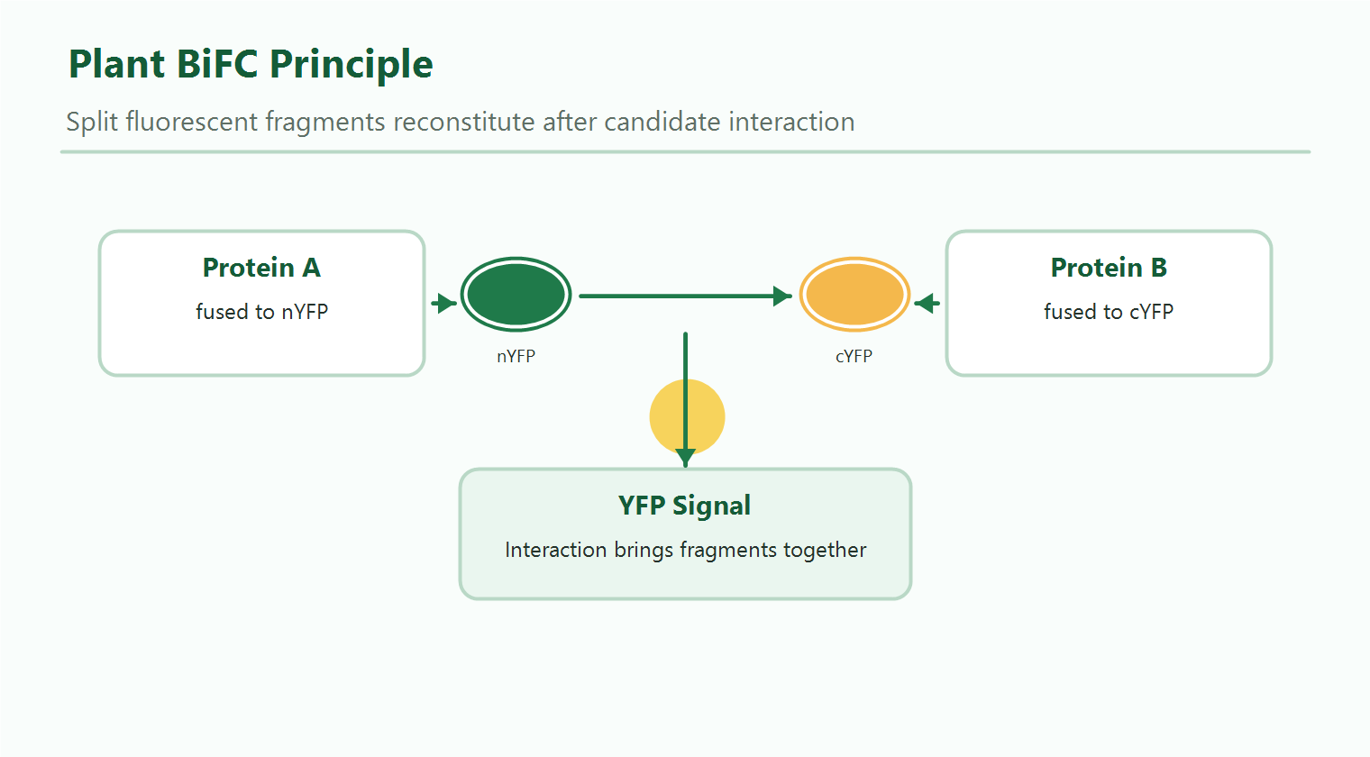
Figure 1. Split fluorescent protein reconstitution in a plant BiFC assay.
Service Scope
Design of nYFP/cYFP or related split-fluorescent protein fusions with attention to reading frame, stop codon, tag position, and protein function.
Gene synthesis, PCR cloning, subcloning, colony screening, plasmid preparation, and sequencing verification can be included according to project needs.
Agrobacterium-mediated transient expression in plant tissue, commonly using Nicotiana benthamiana leaves, can be used for BiFC imaging projects.
YFP, RFP/mCherry marker, bright field/DIC, and merged images can be collected to support interaction and localization interpretation.
Workflow
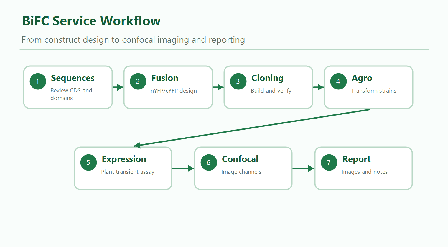
Figure 2. General workflow for plant BiFC service from construct design to confocal imaging.
- Review of candidate protein sequences and project objective
- Fusion orientation and vector design
- BiFC vector construction and sequence verification
- Agrobacterium transformation
- Plant transient expression and timepoint selection
- Confocal imaging of fluorescence and control channels
- Image organization, result interpretation, and report preparation
Recommended Controls
BiFC is strongly dependent on proper controls. Controls help distinguish candidate interaction-associated fluorescence from background complementation, overexpression effects, or non-specific proximity.
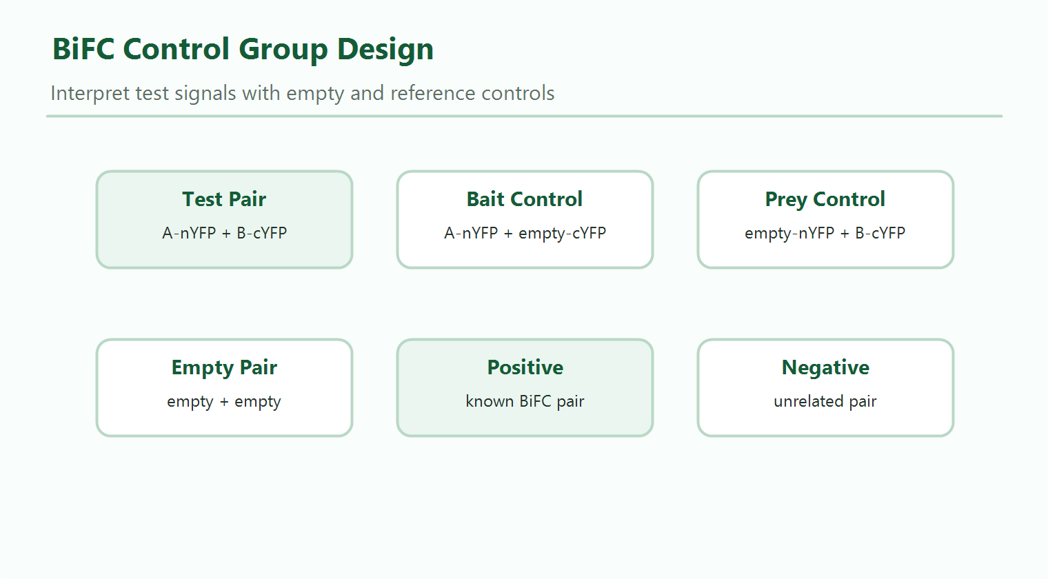
Figure 3. Typical control group design for BiFC interaction testing.
| Control | Purpose |
|---|---|
| Bait fusion + empty complementary fragment | Evaluates background signal from the bait fusion. |
| Prey fusion + empty complementary fragment | Evaluates background signal from the prey fusion. |
| Empty vector pair | Checks system-level background fluorescence. |
| Known positive pair | Confirms that the expression and imaging system is working. |
| Known negative or unrelated pair | Supports specificity interpretation under the same assay conditions. |
Sample and Information Requirements
- Protein or gene names and plant species
- Full CDS or protein sequences
- Known domains, localization motifs, or transmembrane regions
- Preferred fusion orientation, if known
- Existing plasmids, vector maps, resistance information, and sequencing results
- Required localization marker or imaging channel
- Positive or negative control information, if available
- Desired timepoints, number of groups, and image output format
Deliverables
- Construct design summary, when included
- Recombinant vector information and sequencing verification records
- Plasmids or bacterial glycerol stocks, when applicable
- Transient expression and imaging records
- Original confocal images by channel
- Merged representative images
- Control group summary
- Technical report with result interpretation notes
Interpretation Notes
BiFC can provide visual evidence that two candidate proteins are close enough to reconstitute a fluorescent signal in plant cells. However, complementation can be slow and partially irreversible, and signal intensity is affected by construct design, expression level, protein localization, imaging settings, and cellular context. Orthogonal validation such as Co-IP, Y2H, LCI, GST pull-down, or FRET may be recommended for selected protein pairs.
Related Services
For BiFC project design, please contact us with the candidate protein sequences, expected fusion design, and imaging requirements.
For research use only, not for clinical use.